Neural Mobilization Therapy in Queens & Long Island
Your Nerves Move. When They Can't, You Feel It - and Not Just Where the Nerve Is.
Peripheral nerves are not passive cables running through your body. They are living, metabolically active structures that must slide, glide, and elongate through the surrounding tissues with every movement you make. A nerve that can't move freely - because of adhesion, scarring, inflammation, compression, or mechanical restriction - generates pain, numbness, tingling, and weakness that can appear anywhere along its distribution, far from the actual restriction site.
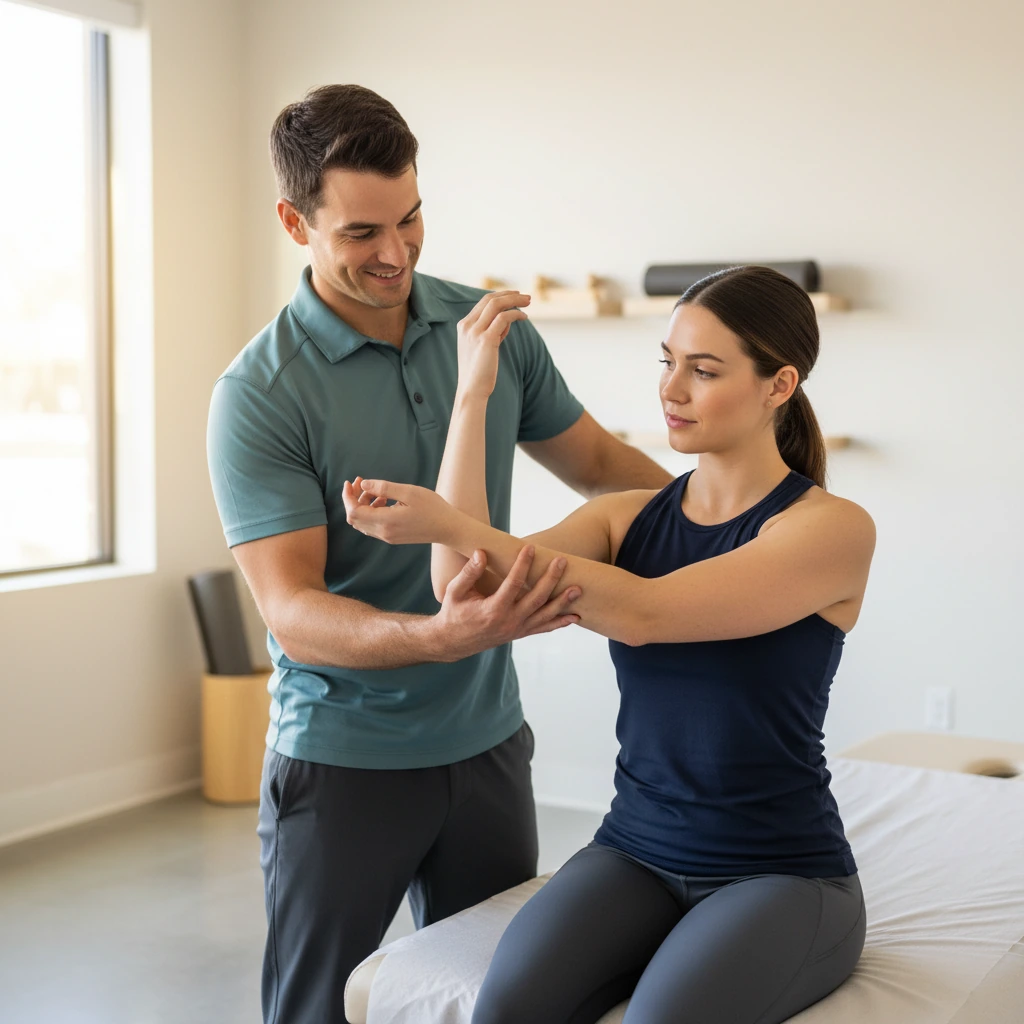
Neural mobilization is a manual and exercise-based physical therapy technique that restores normal nerve movement and extensibility - directly treating the mechanical component of nerve-related pain, radiculopathy, entrapment syndromes, and the referred symptoms they generate. At Dynamic Physical Therapy, neurodynamic assessment and treatment is integrated into management whenever nerve involvement is suspected - using the full range of passive manual techniques, active nerve gliding exercises, and cervical and lumbar neurodynamic mobilization that the evidence supports.
The Mechanics of the Peripheral Nervous System and What Happens When Movement Is Restricted
During normal limb movement, peripheral nerves must travel several centimetres along their path - sliding through adjacent soft tissue, gliding relative to surrounding structures, and elongating under tension as joints move to the end of their range. The median nerve, for example, travels up to 2.4cm longitudinally in the forearm during wrist movement. This movement is not passive; it depends on the mechanical interface between the nerve and its surrounding tissues remaining free of adhesion, compression, and excessive friction.
When nerve movement is restricted - by scarring from surgery or injury, fibrotic adhesion, compression from an adjacent disc or osteophyte, or the cumulative effects of chronic inflammation - the nerve cannot glide freely. The resulting mechanical tension impairs intraneural microcirculation, obstructs axonal transport, creates intraneural oedema, and sensitises the nerve's own pain receptors (nervi nervorum). Neural mobilization directly targets these mechanical restrictions - restoring the slide and glide of the nerve within its tissue bed, reducing intraneural pressure, and improving the intraneural blood flow that nerve health and function require.
Longitudinal Nerve Excursion
Peripheral nerves slide several centimetres longitudinally during normal limb movement. Restriction of this longitudinal excursion - measured with dynamic ultrasound - is the primary mechanical deficit that neural mobilization corrects.
Intraneural Oedema & Microcirculation
Restricted nerve movement impairs the microcirculation within the nerve - reducing the blood flow that delivers nutrients and removes metabolic waste. The resulting intraneural oedema increases nerve sensitivity and impairs signal conduction, producing the pain and neurological symptoms that neural mobilization addresses by restoring circulation through movement.
Mechanical Interface Adhesion
Post-surgical fibrosis, inflammatory adhesion, and chronic compression bind the nerve to adjacent tissue - preventing the independent movement that normal nerve function requires. Neural mobilization breaks down these adhesions through repeated, graded movement at the interface between the nerve and surrounding structures.
Central Sensitisation Modulation
Neural mobilization produces neurophysiological effects beyond the mechanical - activating endogenous pain inhibition pathways, reducing temporal summation of pain signals, and modulating central sensitisation in chronic nerve pain presentations where the peripheral stimulus has long since resolved.
Symptoms That Suggest Neurodynamic Dysfunction
Nerve-related pain has a distinctive pattern - often described differently from musculoskeletal pain, and following distributions that don't match simple muscle or joint pathology. These are the features that suggest the nervous system needs to be assessed and treated.
Nerve-Related Pain Conditions Where Neurodynamic Treatment Produces Meaningful Results
Sciatica & Lumbar Radiculopathy
Cervical Radiculopathy (Arm Pain)
Carpal Tunnel Syndrome
Lateral Epicondylalgia (Tennis Elbow)
Plantar Fasciitis & Tarsal Tunnel
Thoracic Outlet Syndrome
Whiplash-Associated Disorders
Peripheral Neuropathy
Meralgia Paresthetica
Post-Surgical Neural Adhesion
Ulnar Nerve Entrapment (Cubital Tunnel)
Double Crush Syndrome
Neurodynamic Testing - How Neural Involvement Is Confirmed Before Treatment Begins
Neural mobilization begins with neurodynamic assessment - standardised clinical tests that place progressive mechanical tension on specific neural structures and observe whether familiar symptoms are reproduced. A positive neurodynamic test confirms that the nervous system is mechanically sensitised and guides which techniques and nerve tracts require treatment.
Neurodynamic tests are not just diagnostic - they are also prognostic and treatment response indicators. Improvement in the range of movement available before symptoms are provoked, or reduction in symptom intensity during the test, is a direct measure of neural mobilization treatment response session-to-session.
Straight Leg Raise (SLR)
Hip flexion with knee extended - progressively loading the sciatic nerve and its lumbar nerve roots. Positive when familiar leg symptoms are reproduced, distinguished from hamstring tightness by the addition of sensitising movements.
Sciatic nerve / L4 - S1Slump Test
Combined spinal flexion, hip flexion, and knee extension - the most sensitive neurodynamic test for sciatic involvement, loading the neural tissues from the spinal cord through the sciatic nerve in a single, progressive sequence.
Sciatic nerve / dural tissueUpper Limb Neurodynamic Test 1 (ULNT1)
Shoulder depression, abduction, elbow extension, wrist and finger extension - the primary test for median nerve mechanosensitivity and C6/C7 radiculopathy. The cervical lateral glide sensitiser differentiates neural from musculoskeletal origin.
Median nerve / C6 - C7Upper Limb Neurodynamic Test 2b
Shoulder depression, internal rotation, elbow extension, pronation, wrist flexion - biased toward the radial nerve. Positive in lateral epicondylalgia with neural involvement, C5/C6 radiculopathy, and radial tunnel syndrome.
Radial nerve / C5 - C6Prone Knee Bend (PKB)
Prone positioning with passive knee flexion - loading the femoral nerve and L2/L3/L4 nerve roots. The test for anterior thigh pain, groin pain, or upper leg symptoms with a femoral or upper lumbar nerve root component.
Femoral nerve / L2 - L4Upper Limb Neurodynamic Test 3 (ULNT3)
Shoulder depression, abduction, elbow flexion, wrist extension, ulnar deviation - biased toward the ulnar nerve. Used for medial elbow, medial forearm, and ring and little finger symptoms, and cubital tunnel assessment.
Ulnar nerve / C8 - T1What to Expect During Neural Mobilization Treatment
Neurodynamic Assessment
Your therapist performs standardised neurodynamic provocation tests relevant to your symptoms - identifying which neural structures are mechanically sensitised, at which point in the range symptoms are provoked, and whether sensitising manoeuvres (head position, contralateral lateral flexion, ankle dorsiflexion) reproduce your familiar pain pattern. This confirms neural involvement before treatment begins.
Technique Selection - Sliders or Tensioners
Based on the acuity and sensitivity of the nerve presentation, your therapist selects the appropriate technique - slider techniques for acute or sensitised presentations, tensioners for chronic or less irritable cases. The selection is made based on what the nerve can tolerate, not what produces the most intense sensation.
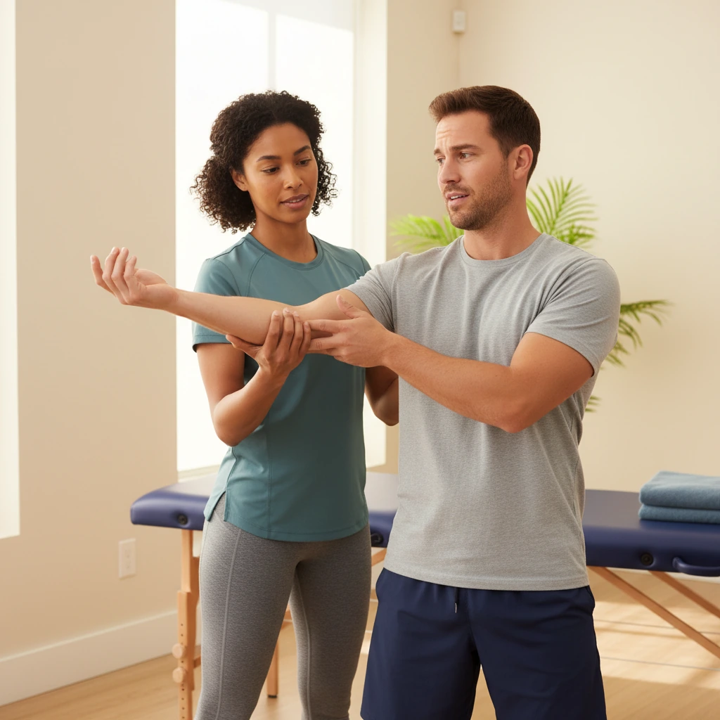
Passive Therapist-Applied Mobilization
Your therapist applies the neurodynamic technique passively - guiding your limb through the sequence of joint positions that produce nerve movement through the restricted mechanical interface. Techniques are applied rhythmically, at a carefully controlled pace, staying within the symptom range that is appropriate for your presentation.
Active Home Exercise Program
Neural mobilization produces its best results with consistent daily repetition. You receive a specific home exercise program of the appropriate slider or tensioner technique for your condition - performed independently between sessions to maintain the nerve movement improvements made in clinic and continue reducing adhesion over time.
Integration with Manual Therapy & Exercise
Neural mobilization is most effective as part of a comprehensive treatment plan that also addresses the mechanical interface - the joints, discs, and soft tissue structures that the nerve passes through. Joint mobilization, myofascial release, and targeted strengthening are integrated alongside neurodynamic treatment for optimal outcomes.

Why Neural Mobilization Should Be Part of Nerve Pain Treatment
Restores Nerve Movement
By directly restoring the longitudinal excursion of restricted nerves through their tissue beds, neural mobilization addresses the mechanical component of nerve entrapment and sensitivity that joint and soft tissue treatment alone cannot reach.
Reduces Intraneural Oedema
Neural mobilization disperses intraneural oedema and improves intraneural microcirculation - reducing the internal pressure that sensitises the nerve's own pain receptors and impairs nerve signal conduction.
Reduces Pain & Neurological Symptoms
Systematic reviews confirm neural mobilization significantly reduces pain, improves function, and reduces neurological symptoms - particularly for cervical radiculopathy, nerve-related low back pain, and plantar heel pain - in populations that are often resistant to standard treatment.
Non-Invasive Alternative
Neural mobilization produces neurophysiological improvements in nerve conduction (confirmed by EMG) and intraneural oedema reduction (confirmed by MRI) - achieving structural change through precise manual technique, without injection, medication, or surgical intervention.
Neural Mobilization FAQs
Is neural mobilization the same as nerve flossing?
"Nerve flossing" is a colloquial term for neural gliding exercises - specifically the slider technique, in which the nerve is moved longitudinally through its tissue bed by alternately lengthening and shortening from each end. Neural mobilization is the broader clinical term that encompasses both slider techniques (nerve gliding/flossing) and tensioner techniques (nerve stretching), as well as the passive therapist-applied neurodynamic mobilization techniques performed in the clinical setting. Consumer-facing nerve flossing videos on YouTube typically describe a version of the slider technique - but without the clinical assessment that determines whether a slider or tensioner is appropriate, which nerve is involved, and at what intensity the technique should be applied, there is significant potential to apply the wrong technique to the wrong nerve at the wrong intensity. At Dynamic PT, neurodynamic exercises are prescribed specifically for your nerve, your presentation, and your current symptom sensitivity.
Does neural mobilization hurt?
Correctly applied neural mobilization should not be painful - though it may reproduce familiar neurological symptoms at the end of the available range. The goal is to work within a comfortable range for slider techniques, and at or near the edge of symptom reproduction for tensioner techniques. Your therapist calibrates the technique intensity to your presentation - acute and sensitised nerves receive slider techniques below the symptom threshold; chronic and less irritable presentations may receive some mild symptom reproduction at end-range tensioner positions. If a technique causes significant pain or a sustained increase in neurological symptoms following the session, the intensity was too high and will be reduced. Minor temporary increase in symptoms after treatment that resolves within 24 hours is normal and not a concern.
How is neural mobilization different from treating a pinched nerve?
A "pinched nerve" typically refers to nerve root compression - from a disc herniation, osteophyte, or foraminal stenosis - which is the structural cause of conditions like sciatica and cervical radiculopathy. Treatment of the compression focuses on decompressing the nerve root - through traction, specific directional exercises (McKenzie method), joint mobilization, or epidural injection. Neural mobilization addresses the mechanical component of nerve movement through the surrounding tissue bed - distinct from, but complementary to, treating the compression itself. In practice, both components often need to be addressed: the compression reduces the space available to the nerve, and the restricted nerve movement amplifies the effect of that compression. At Dynamic PT, both are assessed and treated as part of a comprehensive plan.
Can neural mobilization help carpal tunnel syndrome without surgery?
For mild to moderate carpal tunnel syndrome - confirmed with in-clinic EMG/NCS testing - neural mobilization has strong evidence of producing meaningful clinical improvement. Multiple RCTs demonstrate reduction in median nerve latency (the EMG measure of nerve compression severity), reduction in intraneural oedema confirmed on MRI, and significant improvements in pain, grip strength, and hand function. Neural mobilization is most effective for CTS when combined with carpal bone mobilization, nerve and tendon gliding exercises, and splinting - and when applied early in the condition before structural nerve damage becomes advanced. For patients with severe CTS or significant thenar muscle wasting, surgical decompression may still be the most appropriate intervention - your therapist will give you an honest assessment of whether conservative management is likely to be sufficient for your specific severity.
Is neural mobilization covered by insurance?
Neural mobilization delivered as part of a physical therapy plan is covered by Medicare, Medicaid, and most commercial insurance plans - billed as manual therapy or therapeutic exercise within your PT session. At Dynamic Physical Therapy, we verify your complete benefits before your first appointment and perform EMG/NCS nerve testing in-clinic on the same day as your assessment, which is separately covered under diagnostic testing benefits. Call (516) 661-9880 and our team will confirm your coverage.