Musculoskeletal Ultrasound (MSKUS) in Queens & Long Island

Real-Time Imaging of the Structures Behind Your Pain - In the Clinic, During Your Visit
Musculoskeletal ultrasound (MSKUS) uses high-frequency sound waves to produce real-time, high-resolution images of muscles, tendons, ligaments, bursae, nerves, joints, and cartilage - without radiation, without contrast injections, and without the enclosed environment of an MRI scanner. It is the fastest-growing diagnostic imaging modality in musculoskeletal medicine, and for good reason: it shows the tissue while it moves.
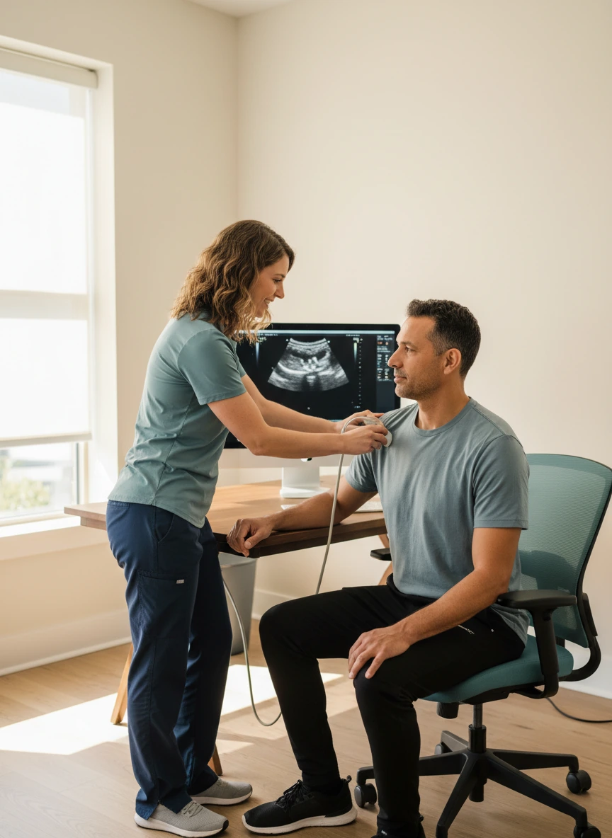
At Dynamic Physical Therapy, MSKUS is performed in-clinic by our trained therapists as a point-of-care diagnostic tool - conducted during your assessment or treatment visit, with findings reviewed immediately and integrated directly into your clinical diagnosis and rehabilitation plan. No referral to an imaging centre. No days of waiting for a report. The image is produced, interpreted, and applied to your care in the same session - which is the defining clinical advantage of having diagnostic ultrasound within a physical therapy practice.
How Musculoskeletal Ultrasound Works - and Why Frequency Matters
Musculoskeletal ultrasound uses a handheld transducer pressed against the skin that emits high-frequency sound waves - typically 7 to 17 MHz for superficial musculoskeletal structures. These sound waves travel through soft tissue and are reflected back at different rates depending on the density and acoustic properties of each tissue type. The transducer captures the returning echoes and a computer converts them into a real-time image displayed on the monitor.
Different tissues have characteristic ultrasound appearances. Healthy tendon has a bright, fibrillar (striated) pattern; tendinopathy appears darker and loses the fibrillar structure. Fluid is black (anechoic); blood vessels pulsate with Doppler colour. Nerves have a fascicular honeycomb appearance. These recognisable tissue signatures, combined with the ability to move the probe and ask the patient to perform movements during imaging, make MSKUS uniquely suited to soft tissue diagnosis in a clinical setting.
Tendons
MSKUS's strongest application. Healthy tendon shows bright, organised fibrillar echoes; tendinopathy shows focal hypoechoic areas of collagen disruption; full-thickness tears show complete discontinuity. MSKUS matches MRI accuracy for rotator cuff, Achilles, patellar, and posterior tibial tendon assessment.
Muscles
Muscle tears show as hypoechoic defects within the muscle belly; haematoma appears as a fluid collection; chronic denervated muscle shows increased echogenicity. Dynamic assessment directly visualises muscle contraction, activation, and sliding between muscle layers during movement.
Bursae & Fluid Collections
Fluid reliably appears as anechoic (black) areas - making joint effusions, bursal swelling, Baker's cysts, ganglion cysts, and haematomas immediately identifiable and measurable. Power Doppler detects active synovial inflammation even without visible fluid by identifying hyperaemia.
Nerves & Ligaments
Peripheral nerves can be traced along their course and assessed for swelling (increased cross-sectional area), loss of fascicular pattern, or compression at entrapment sites. Ligaments show as bright, fibrillar bands at joint margins; partial or complete tears disrupt this pattern and may show surrounding fluid.
What MSKUS Can Diagnose That Static MRI Cannot - Real-Time Dynamic Imaging
The defining clinical superiority of musculoskeletal ultrasound over MRI for specific conditions is its ability to image structures while they move. Many musculoskeletal pathologies are only apparent during specific movements - and simply do not exist on a static MRI scan taken with the patient lying still.
Shoulder Impingement - During Abduction
Subacromial impingement is a dynamic event - the rotator cuff and bursa compress under the acromion during shoulder elevation, not at rest. MSKUS directly visualises the moment of impingement during active arm elevation, showing exactly which structures are compressed. Static MRI shows the anatomy but cannot demonstrate the dynamic compression.
Only visible during movementTendon Subluxation & Snapping
Biceps long head tendon subluxation, extensor carpi ulnaris subluxation, and peroneal tendon subluxation all produce symptoms only during specific movements. On static MRI, the tendon is often back in its correct groove. Dynamic MSKUS during the provocative movement directly visualises the tendon jumping out of its groove - confirming a diagnosis MRI would miss entirely.
Invisible on static MRISnapping Hip Syndrome
The external snapping hip - produced by the iliotibial band or gluteus maximus snapping over the greater trochanter - is a purely dynamic phenomenon. MSKUS during the provocative hip movement directly demonstrates the snap, identifies the structure involved, and rules out intra-articular causes. MRI of a snapping hip is almost universally normal.
Requires dynamic imagingTrigger Finger - Tendon Catching
The mechanical catching of the flexor tendon at the A1 pulley in trigger finger can be directly visualised by MSKUS during active finger flexion and extension - showing the nodule catching on the pulley, the degree of restriction, and the severity of the condition in real time. This guides conservative management decisions and monitors treatment response.
Real-time catching visibleMuscle Herniation Through Fascia
Transfascial muscle herniation - where muscle herniates through a defect in the deep fascia during contraction - is only present when the muscle contracts. At rest, the hernia reduces and the fascia looks intact. Dynamic MSKUS during muscle contraction reveals the defect and the herniation that disappears at rest - consistently missed by static MRI.
Only present during contractionJoint Instability - Stress Testing
Ultrasound can be performed during manual stress testing of ligaments - directly visualising the gap width at the joint line under valgus or varus stress. This quantifies ligament laxity in real time, distinguishing a structurally intact but clinically lax ligament from a functionally significant partial tear that static MRI under no stress cannot provide.
Stress testing in real timeClinical Situations Where MSKUS Provides Immediate Diagnostic Value
MSKUS adds most value when clinical examination has identified a suspected soft tissue pathology that needs immediate visual confirmation - or when a dynamic component to the symptoms needs to be demonstrated that static imaging cannot capture.

Musculoskeletal Conditions Evaluated with MSKUS at Dynamic PT
Rotator Cuff Tears & Tendinopathy
Achilles Tendinopathy & Tears
Patellar Tendinopathy
Bursitis (Subacromial, Trochanteric, Olecranon)
Carpal Tunnel & Nerve Entrapment
Lateral Epicondylitis (Tennis Elbow)
Biceps Tendon Pathology
Hamstring & Muscle Tears
Ganglion Cysts & Soft Tissue Masses
Snapping Hip & Shoulder Syndrome
Trigger Finger (Flexor Tendon)
Joint Effusion & Synovitis
MSKUS vs. MRI - When Each Is the Better Choice
MSKUS and MRI are complementary tools - each superior for different clinical questions. Understanding when to use each is what clinical experience with both modalities provides.
MSKUS Is Preferred When…
The question involves dynamic soft tissue assessment, immediate point-of-care diagnosis, or structures that move during symptoms.
MRI Is Preferred When…
The question involves deep intra-articular structures, bone marrow, cartilage, or the internal architecture of joints inaccessible to sound waves.
What to Expect During Your Musculoskeletal Ultrasound at Dynamic PT
No Special Preparation Required
Unlike MRI, MSKUS requires almost no preparation. Wear comfortable clothing that allows access to the area being examined. No fasting, no contrast injections, no removal of metallic implants. If you have a joint replacement, pacemaker, or spinal hardware, MSKUS is completely unaffected - and may be the preferred imaging choice precisely because of those implants.
Transducer Application & Gel
A small amount of water-based ultrasound gel is applied to the skin over the area being examined - this creates the acoustic coupling that allows sound waves to enter the tissue without air gaps. The gel is colourless, water soluble, and wipes off completely. The transducer is pressed gently against the skin and moved over the area of interest.
Static and Dynamic Assessment
Your therapist examines the target structures in longitudinal and transverse orientations. You will then be asked to perform specific movements during imaging - elevating your arm, bending your knee, flexing your fingers. These dynamic views reveal pathologies that only appear during movement, and are the most clinically valuable component of many MSKUS examinations.
Bilateral Comparison When Indicated
One of MSKUS's unique advantages is the ability to immediately compare the symptomatic side to the asymptomatic side in the same session without scheduling a separate examination. Many measurements - tendon thickness, nerve cross-sectional area, bursal fluid volume - are most accurately interpreted by comparison to the contralateral normal side.
Immediate Results & Treatment Integration
Your therapist discusses the findings with you immediately after imaging - showing you the images, explaining what the ultrasound revealed, and describing how the findings change or confirm the diagnosis and treatment approach. No waiting for a radiology report. The imaging finding is applied to your care in the same session it was acquired.
The Four Clinical Advantages of In-Clinic MSKUS
Real-Time Dynamic Imaging
The only imaging modality that shows soft tissue pathology during the movement that provokes symptoms - directly visualising tendon subluxation, snapping, impingement, and muscle herniation that static MRI cannot capture.
No Radiation, No Contrast
Completely safe for all patient populations - pregnant patients, children, patients with renal impairment who cannot receive contrast agents, and patients requiring frequent serial imaging to monitor treatment response. No known adverse effects at diagnostic intensities.
Point-of-Care, Same-Visit Results
Performed during your PT visit. Findings reviewed immediately. Treatment decisions made that day. No referral pathway, no imaging centre appointment, no waiting days or weeks for a report. The clinical bottleneck of the diagnostic wait is removed entirely.
Serial Monitoring Without Burden
Unlike MRI, MSKUS can be repeated as many times as clinically indicated without radiation concern, significant cost, or scheduling delays. Serial imaging at defined intervals objectively tracks healing - providing the evidence base for progression decisions that clinical examination alone cannot.
Musculoskeletal Ultrasound FAQs
Is musculoskeletal ultrasound as accurate as MRI?
For many specific soft tissue conditions, MSKUS has been shown to be equivalent to or better than MRI. Published evidence confirms MSKUS equals MRI accuracy for rotator cuff tear identification, Achilles tendinopathy, lateral epicondylitis, peripheral nerve injuries, patellar tendon pathology, and thumb ulnar collateral ligament tears. For deep intra-articular structures - cruciate ligaments, labrum, meniscal tears - MRI remains superior because ultrasound cannot penetrate the bone surfaces surrounding joint cavities. The appropriate question is not "which is better overall?" but "which is better for this specific clinical question?" - and for the majority of soft tissue pathologies assessed in physical therapy, MSKUS performs at least comparably to MRI, with the additional advantage of dynamic imaging capability.
Can I have MSKUS if I have a joint replacement or metal implant?
Yes - and in many cases, MSKUS is the preferred imaging modality precisely because of metallic implants. MRI is significantly degraded by metallic hardware - rods, screws, prostheses, and implants create artefacts that obscure surrounding soft tissue and can make the study non-diagnostic. MSKUS is completely unaffected by metallic implants - the sound waves travel around and between the metal surfaces without any interference. For patients with hip or knee replacements, spinal fusion hardware, or any other metallic implant, MSKUS is often the only soft tissue imaging option that provides usable results in the vicinity of the implant.
Is MSKUS painful?
Diagnostic MSKUS is painless - the transducer is pressed gently against the skin with ultrasound gel. Most patients find the gel slightly cool but otherwise feel nothing from the imaging itself. If the area being examined is already tender, the light pressure of the transducer may cause mild discomfort over a particularly inflamed or sensitive spot - but this is incidental to the examination, not caused by any aspect of the ultrasound technology. The transducer pressure is adjusted to minimise discomfort over tender areas while still obtaining diagnostic images.
What is the difference between diagnostic and therapeutic ultrasound?
These are completely different modalities that share only the name "ultrasound." Diagnostic musculoskeletal ultrasound (MSKUS) uses high-frequency sound waves (7 - 17 MHz) at very low intensity to produce real-time images of tissue - it produces no heating and has no therapeutic effect. It is purely an imaging tool. Therapeutic ultrasound - often applied with a round applicator in physiotherapy for pain relief and tissue heating - uses much lower frequencies (1 - 3 MHz) at higher intensities to produce thermal and non-thermal effects in tissue. At Dynamic PT, diagnostic MSKUS is the point-of-care imaging modality; therapeutic ultrasound is a separate treatment modality used for different clinical indications.
Is musculoskeletal ultrasound covered by insurance?
MSKUS coverage varies by plan and clinical indication. When performed as a diagnostic imaging study for a specific musculoskeletal complaint, it is covered by Medicare, Medicaid, Workers' Compensation, No-Fault, and many commercial insurance plans under diagnostic ultrasound procedure codes. No-Fault insurance (motor vehicle accidents) frequently covers MSKUS as part of the diagnostic work-up for soft tissue injuries. At Dynamic Physical Therapy, we verify your specific coverage before your appointment. Call (516) 661-9880 and our team will confirm how your plan covers diagnostic ultrasound.